Sleep is a fundamental biological process that significantly influences cognitive function, emotional well-being, and overall health. It is regulated by a complex interaction of neurological and hormonal mechanisms that help align the body’s internal clock, known as the circadian rhythm. An important part of this interaction is the small structure deep within the brain called the pineal gland.
Understanding pineal gland function is essential for understanding how the brain controls the sleep–wake cycle. So, what does the pineal gland do? Despite its modest size, the pineal gland plays a vital role in managing the sleep–wake cycle through the secretion of melatonin, a hormone involved in signalling the onset of sleep. As the main source of melatonin, the pineal gland plays a central role in maintaining circadian rhythm and supporting healthy sleep. Disruptions in this rhythm are linked to common sleep disorders such as insomnia, jet lag, and delayed sleep-wake phase disorder (DSWPD). This article will discuss the anatomy, function, and clinical significance of the pineal gland and its role in melatonin production.
What does the pineal gland do?
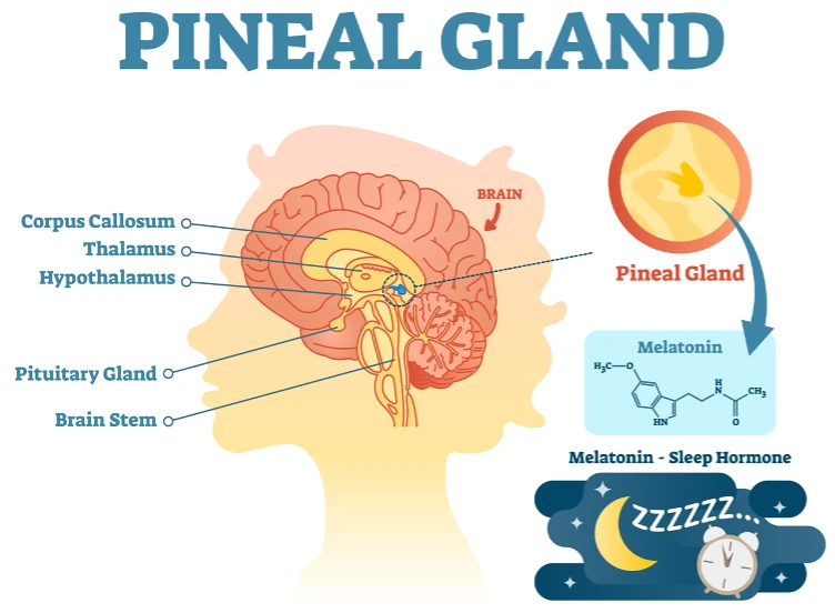
The pineal gland is a small, pinecone-shaped structure situated near the centre of the brain, between the two hemispheres. Typically measuring around 5 to 8 mm in length, it is considered a component of the endocrine system due to its ability to secrete hormones directly into the bloodstream. The core function of the pineal gland is the production and regulation of melatonin. Melatonin secretion is intimately tied to the body’s perception of light and darkness. In response to decreasing ambient light, detected by the retina and relayed through the suprachiasmatic nucleus (SCN) of the hypothalamus, the pineal gland increases melatonin production, signalling the body that it is time to sleep. Conversely, exposure to light inhibits melatonin synthesis, promoting alertness and wakefulness.
Beyond its role in melatonin synthesis, the pineal gland serves as a biological timekeeper, helping to regulate circadian rhythms, internal cycles that influence sleep, hormone secretion, appetite, and body temperature. The pineal gland also has a secondary influence on reproductive hormones, particularly in seasonal breeders in the animal kingdom, although the significance of these findings in humans remains under investigation.
Pineal gland function in sleep and health
The pineal gland helps to regulate sleep mainly by producing melatonin. Melatonin levels start to increase in the evening, reach their highest point at night, and drop by morning. This daily pattern helps the body feel sleepy at night and alert during the day. When this rhythm runs smoothly, it supports good sleep, clear thinking, and a stable mood.
When the pineal gland function is impaired due to ageing, lifestyle factors, or neurological conditions, the consequences can manifest as disrupted sleep onset, poor sleep quality, or reduced sleep duration. Insufficient melatonin secretion has been linked to several sleep disorders, including insomnia and circadian rhythm sleep–wake disorders such as delayed sleep-wake phase disorder. Disruption of the pineal gland’s function may also have broader health implications. Poor-quality or inadequate sleep caused by pineal gland dysfunction can weaken the immune system, increase the risk of metabolic conditions like obesity and type 2 diabetes, and negatively affect mental health.
How is melatonin produced?
To understand how melatonin regulates sleep, it is essential to explore how it is synthesised. Melatonin is derived from serotonin, a neurotransmitter that originates from the amino acid tryptophan. This multi-step biochemical process begins with the dietary intake of tryptophan, which is converted into serotonin within the brain. In the pineal gland, serotonin undergoes enzymatic transformation into melatonin.
Melatonin production is closely controlled by exposure to light. When the eyes detect light, signals are sent through a special pathway to the brain’s master body clock, called the suprachiasmatic nucleus (SCN). The SCN then sends messages through several parts of the nervous system to the pineal gland. In darkness, this pathway tells the body to produce the pineal gland hormone (melatonin). In contrast, exposure to light stops melatonin production, helping the body stay awake and alert.
This natural cycle makes sure that melatonin levels go up in the evening to help you fall asleep and go down in the morning to help you wake up. Melatonin works like a signal to tell the body it’s night-time, helping your internal clock match the outside world’s light and dark pattern. Knowing how melatonin is produced helps us understand how sleep works and is useful in finding and treating sleep–wake cycle disorders.
Pineal gland hormones and their roles
Melatonin is the primary hormone secreted by the pineal gland, and it exerts systemic effects far beyond its role in sleep regulation. It possesses potent antioxidant properties, neutralising free radicals and reducing oxidative stress in various tissues. This has led to interest in melatonin’s potential protective role in neurodegenerative disorders, cardiovascular health, and cancer prevention, although conclusive evidence in humans remains limited.
The pineal gland may also secrete other bioactive compounds, such as peptides and neurotransmitters, though their roles are not fully understood. Some researchers have suggested that the pineal gland may influence reproductive hormone regulation. In particular, melatonin is thought to affect the timing of puberty and reproductive cycles, especially in seasonal breeders. The systemic effects of melatonin include modulation of mood, regulation of immune responses, and interaction with other hormonal systems. These many roles show that melatonin is more than just a sleep aid; it is a hormone that affects many important functions in the body.
Factors affecting pineal gland function
A variety of factors can influence pineal gland function and subsequently disrupt melatonin production. One of the most significant factors is light exposure, particularly blue light emitted by electronic devices such as smartphones, tablets, and computer screens. Evening exposure to artificial light can suppress melatonin secretion, delaying sleep onset and reducing overall sleep quality. Shift work and irregular sleep schedules also challenge the natural circadian rhythm, leading to mismatches between internal biological timing and external demands.
Ageing is another factor; as individuals grow older, melatonin production tends to decline, potentially contributing to more fragmented and less restorative sleep in older adults. Certain medications, including beta-blockers, non-steroidal anti-inflammatory drugs (NSAIDs), and some antidepressants, may interfere with melatonin synthesis or secretion. Medical conditions such as tumours of the pineal gland or neurological disorders affecting this neurological pathway can also impair function.
In some cases, exogenous melatonin supplements may be recommended to address deficiencies or to assist in the treatment of circadian rhythm disorders. When used appropriately and under medical supervision, melatonin supplementation can improve sleep latency and duration, particularly in cases of jet lag, shift work sleep disorder, and delayed sleep-wake phase disorder (DSWPD).
Understanding the pineal gland’s role in sleep: final thoughts
The pineal gland plays an important role in regulating the body’s internal clock through its secretion of melatonin, a hormone that governs the timing, duration, and quality of sleep. Understanding pineal gland function offers valuable insight into the physiological basis of sleep and provides a foundation for addressing a range of sleep disorders. For both clinicians and individuals, exploring the mechanisms by which the pineal gland influences sleep can lead to more effective management of insomnia and other sleep-related issues.
- If you are experiencing persistent difficulty falling asleep, staying asleep, or waking too early, these issues may be linked to disruptions in melatonin production or pineal gland function.
- Addressing this imbalance can lead to significant improvements in sleep quality, energy levels, and overall well-being.
- Effective treatment can have wide-ranging benefits, not only enhancing restfulness but also improving concentration and emotional stability in both personal and professional life.
- Better sleep contributes to improved heart health, stronger immune function, and a more balanced hormonal system.
Quality sleep is achievable with the right support. If you’re seeking a personalised sleep assessment, expert guidance on melatonin, or help to enhance your sleep quality, a tailored approach could make all the difference. Schedule a consultation to explore your concerns and receive advice that fits your specific needs. Addressing sleep issues effectively can lead to sharper focus, a more balanced mood, and a clear improvement in your overall health.